Advanced Digital Imaging for Oral and Maxillofacial Surgery
in Scottsdale, AZ
What Is Digital Imaging in Oral and Maxillofacial Surgery?
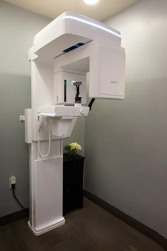
At The Pogue Center, we use advanced digital imaging technology to plan and perform oral and maxillofacial surgery with unmatched precision and safety. Our state-of-the-art equipment helps us get a clear, 3D view of your anatomy—right down to the root.
This isn’t just better technology—it means better outcomes, faster healing, and more comfortable care for you. Our state-of-the-art technology helps your board-certified oral and maxillofacial surgeon make confident, informed decisions to protect your health and smile.
Schedule a Consultation
Oral & Maxillofacial Surgery Digital Imaging Terms to Know
In oral and maxillofacial surgery, digital imaging plays a crucial role in diagnosis, treatment planning, and surgical precision. To help you talk with your surgeon during your consultation, here are some common imaging terms to know.
- Low-radiation imaging refers to advanced imaging technologies, like digital X-rays and CBCT scans, that use much less radiation compared to traditional X-rays. This makes them safer for patients while still providing highly detailed images for accurate diagnosis and treatment planning. It’s a key benefit, especially when multiple scans are needed for procedures like implants or extractions.
- Cone Beam CT Scan (CBCT), also called 3D Cone Beam Imaging, is a special type of 3D X-ray that takes detailed pictures of your teeth, jaw, and surrounding structures. It provides a complete, 3-dimensional view, helping your oral surgeon plan treatments like dental implants, extractions, or jaw surgery with great precision and safety. CBCT scans offer more detailed information than regular X-rays, so your surgeon can avoid nerves and other important areas during procedures.
- Digital X-rays: Standard 2D images for evaluating tooth structure, bone levels, and detecting issues like impacted teeth or infections.
- Intraoral Cameras: Small cameras used to capture detailed images inside the mouth, helping both patients and surgeons visualize problem areas.
- Panoramic X-rays (Full Mouth X-ray): A broad view of the entire mouth, jaws, and surrounding structures, often used for initial assessment.
- Dental X-rays: Traditional or digital images of your teeth and jaw used to detect cavities, bone loss, impacted teeth, and other dental issues.
- Digital X-rays: Modern dental X-rays captured with digital sensors instead of film. They provide faster results, lower radiation exposure, and enhanced image quality.
- Digital Scan: A broader term that often refers to intraoral scans—3D digital impressions of your teeth and gums using a small wand-like scanner. These scans are used for precise treatment planning, such as creating crowns, implants, or aligners.
- A 3D dental scan is a special digital image that creates a detailed, three-dimensional picture of your teeth, gums, and jaw. It helps your oral surgeon see everything clearly to plan your treatment more accurately. This means safer procedures and better results for you.
These imaging technologies allow oral surgeons to plan procedures with higher accuracy, reduce risks, and improve outcomes.
Symptoms or Conditions That May Require Digital Imaging and Oral and Maxillofacial Surgery:
- Severe Tooth Pain: Especially when caused by impacted or infected teeth (like wisdom teeth).
- Swelling in the Jaw, Face, or Gums: Can signal infection, cysts, or hidden abscesses that need evaluation.
- Chronic Sinus Pressure or Discomfort Near the Upper Jaw: May indicate sinus complications related to oral health.
- Difficulty Opening the Mouth Fully: Could be related to jaw joint (TMJ) disorders or muscle/bone abnormalities.
- Loose or Missing Teeth: Often a sign of bone loss or gum disease; may require dental implants or grafting.
- Jaw Pain or Popping: May suggest joint misalignment, trauma, or structural issues.
- Visible Jaw Misalignment or Bite Problems: Orthognathic (jaw) surgery planning typically requires detailed 3D imaging.
- Facial Trauma or Injury: Broken or displaced facial bones must be precisely imaged for surgical repair.
- Persistent Infections Around the Teeth or Gums: These could point to underlying bone damage or failed previous treatments.
- Numbness or Tingling in the Lips, Chin, or Face: May indicate nerve involvement needing careful mapping with imaging.
- Bulging or Abnormal Growth in the Mouth or Jaw: Could be a tumor, cyst, or other lesion that requires biopsy and surgical removal.
- Preparation for Dental Implants: Imaging is essential to assess bone quality, height, and nerve locations.
- Impacted Teeth (Teeth That Haven’t Erupted Properly): Especially common with wisdom teeth and some canine teeth.
- Frequent Headaches or Earaches Tied to Jaw Function: Could stem from jaw joint problems or misalignment.
- Snoring or Breathing Issues During Sleep: Imaging may be needed for patients being evaluated for sleep apnea surgery.
These symptoms often signal underlying issues that require precise diagnosis and treatment planning, which is exactly what digital imaging technology enables at The Pogue Center.
Conditions That May Require Digital Imaging for Oral and Maxillofacial Surgery:
- Impacted Teeth: Teeth that are trapped beneath the gums or bone (commonly wisdom teeth or canines).
- Tooth and Jaw Infections: Including abscesses, osteomyelitis (bone infection), or periapical infections.
- Jaw Misalignment (Malocclusion): Conditions like underbite, overbite, or open bite often require jaw surgery and 3D imaging.
- Temporomandibular Joint Disorder (TMJ/TMD): Imaging helps assess joint damage or misalignment for surgical or non-surgical treatment.
- Cysts and Tumors of the Jaw: Including benign growths like odontogenic cysts and more serious lesions.
- Facial Trauma or Fractures: Any injury involving broken or displaced bones in the jaw, cheek, or eye socket.
- Severe Periodontal Disease With Bone Loss: May require bone grafting and implant planning.
- Sleep Apnea (Obstructive): In some cases, surgical correction of airway obstructions is planned with imaging.
- Missing Teeth Requiring Dental Implants: Bone structure, nerve pathways, and sinus positioning must be mapped precisely.
- Cleft Lip and Palate or Congenital Jaw Deformities: Complex craniofacial anomalies require imaging for surgical planning.
- Oral Pathology: Any suspected abnormality in the mouth, jaw, or salivary glands that needs biopsy or surgical removal.
- Jaw Cysts or Bone Lesions: Imaging is critical for identifying the extent and location before surgical intervention.
- Osteonecrosis of the Jaw (ONJ): Bone death is often linked to medications like bisphosphonates or radiation therapy.
- Ankylosis (Jaw Joint Fusion): A rare condition where the jaw joint becomes stiff or immobile, often requiring surgical correction.
- Failed Previous Dental Surgeries or Implants: Digital imaging helps assess complications and guide revision surgery.
These conditions require the expertise of a board-certified oral and maxillofacial surgeon—and high-quality digital imaging is often the first step toward safe, successful treatment.

Empowering Patients Through Education: Informed Consent and Oral and Maxillofacial Surgery Videos
At The Pogue Center, we’re dedicated to providing the best oral and maxillofacial surgery experience, and part of that means upholding the highest ethical standards. As part of this commitment, we offer a comprehensive Patient Library.
In our Patient Library, you will find special videos designed to promote transparency, reduce anxiety, and ensure every patient feels confident and well-informed before treatment.
- Informed Consent Videos help patients understand the risks and benefits.
- General Education videos and our other resources help patients fully understand the procedures, the preparation, and the recovery phases.